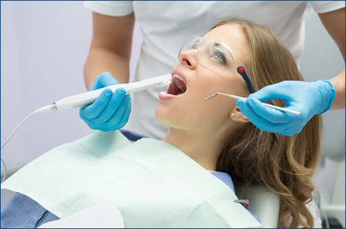
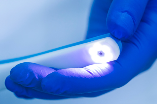
Intraoral photography has become an invaluable tool in modern dentistry, offering detailed visual documentation of a patient’s oral condition. These high-resolution images provide both dentists and patients with a clearer understanding of oral health issues, enhancing diagnosis and treatment planning. By capturing images of the teeth and soft tissues, intraoral photos facilitate better communication and more effective preventive dental care.
 Enhanced Diagnosis
-
- Detailed Visualization: Intraoral photos provide dentists with detailed images of the teeth, gums, and oral tissues. Unlike X-rays, which show bone structure, intraoral photos reveal surface details such as plaque, decay, cracks, and wear patterns. This level of detail helps dentists identify potential problems early, allowing for timely intervention and treatment.
- Comprehensive Record Keeping: By documenting the initial condition of a patient’s mouth, intraoral photos create a comprehensive record that can be referred to over time. This is particularly useful for tracking the progression of dental issues, assessing the effectiveness of treatments, and making informed decisions about future care. Dentists can compare images taken at different appointments to monitor changes and adjust treatment plans as needed.
Improved Patient Communication
-
- Visual Explanations: Intraoral photos enable dentists to show patients exactly what is happening in their mouths. Seeing an image of a cavity or gum disease can be more persuasive and understandable than verbal explanations alone. Patients can see the issues firsthand, making them more likely to understand the need for treatment and comply with recommended care plans.
- Educational Tool: These images serve as an educational tool, helping patients understand their oral health better. Dentists can use intraoral photos to illustrate the importance of good oral hygiene, demonstrate the impact of certain habits, and explain how treatments will address specific problems. This enhances patient engagement and encourages proactive participation in their oral health care.
Precision in Treatment Planning
-
- Accurate Treatment Planning: Intraoral photos provide a clear and precise view of a patient’s oral condition, enabling dentists to develop more accurate and tailored treatment plans. Whether it’s planning a restorative procedure, designing orthodontic treatment, or preparing for cosmetic enhancements, these photos help ensure that the treatment approach aligns closely with the patient’s specific needs.
- Enhanced Collaboration: When collaborating with specialists or other healthcare providers, intraoral photos offer a clear visual reference that aids in discussing complex cases. These images can be easily shared digitally, allowing for more efficient and coordinated care between professionals.
Intraoral photography is a powerful tool that enhances the quality of dental care by improving diagnosis, patient communication, and treatment planning. By providing detailed and accurate images of a patient’s oral condition, intraoral photos support more effective and personalized care. They help dentists make informed decisions, engage patients in their oral health, and facilitate collaboration with other professionals. As technology continues to advance, the use of intraoral photography will likely become even more integral to achieving optimal dental outcomes and patient satisfaction.


